trons surround atoms more or less uniformly,
31
Information about Using Technology to Study Cellular and Molecular Biology
Using Technology to Study Cellular and Molecular Biology
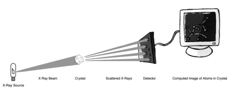
Figure 12. The X-ray crystallography process.
it is possible to determine where atoms are
One of the most striking advancements has
located by looking at these maps. By rotating
been the use of synchrotron X-rays, which are
the crystal and generating an electron density
produced by the bending of particle beams gen-
map for each angle of rotation, it is possible to
erated by large accelerators. In a synchrotron,
produce a three-dimensional model of the mol-
charged particles, such as electrons or posi-
ecule. If the amino acid sequence of a protein
trons, are orbited around a path nearly a mile
is known, an accurate model of the protein can
in circumference, which must be maintained
be generated by fitting the atoms of the known
in a vacuum. Understandably, synchrotrons are
sequence into the electron density map.
quite expensive to build and to maintain, and
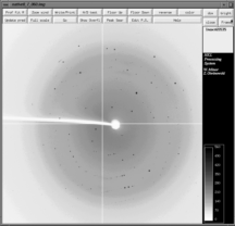
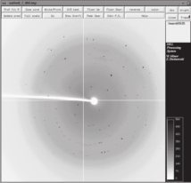
Figure 13 shows a typical diffraction pattern for
a single orientation of a protein crystal through
which an X-ray beam has been passed. Note
the different positions and intensities of the
spots, which mark the locations where scattered
X-rays have struck the detector. The image is
divided into quadrants because the detector was
composed of four separate, adjacent modules.
The white circle to the right of center with the
white line extending to the left is a shadow
resulting from a “beamstop.” The beamstop is a
small piece of lead mounted on a metal arm. It
prevents the intense beam of unscattered X-rays
from impinging on and damaging the detector.
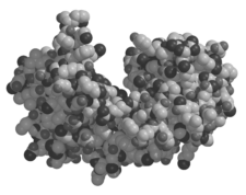
Figure 14 shows a three-dimensional model of a
protein that was crystallized and then analyzed
by X-ray crystallography.
Figure 13. A typical X-ray−diffraction pattern for
a single orientation of a protein crystal through
Equipment used in X-ray crystallography con-
which an X-ray beam has been passed.
tinues to undergo development and refinement.
32
synchrotron radiation. A modern synchrotron
source can reduce total data collection to just
30 minutes, as compared with weeks using ear-
lier X-ray–diffraction equipment.
Determining structures by X-ray diffraction
continues to add to our understanding of DNA
replication and protein synthesis. For example,
scientists recently studied the crystal structures
of a bacterial DNA polymerase I that had DNA
primer templates bound to its active site.13
The enzyme was catalytically active, which
allowed for direct observation of the products
of several rounds of nucleotide incorporation.
The polymerase was able to retain its ability to
Figure 14. Three-dimensional structure of the
distinguish between correctly and incorrectly
DNA-repair protein MutY as determined by X-ray
paired nucleotides in the crystal. By comparing
crystallography. Graphic was produced from infor-
the structures of successive complexes, it was
mation available at http://www.rcsb.org/pdb/ .
possible to determine the structural basis for
sequence-independent recognition of correctly
formed base pairs.13
there are fewer than 20 in the world. Because
synchrotron X-ray beams are many orders of
Ribosomes are the largest asymmetric structures
magnitude brighter than the usual laboratory X-
to be solved by X-ray crystallography so far.
ray sources, data for single crystal orientations
Results, with resolutions as high as 2.4 Å, have
can be collected with exposures of a minute or
helped establish the locations of the 27 proteins
less, rather than exposures of several minutes to
and the 2,833 bases of ribosomal (rRNA) found
an hour.
within the ribosome.4 The structure also shows
that contacts between the two ribosome sub-
The completion of the Human Genome Proj-
units are limited, which helps explain why the
ect has provided the foundation for explosive
ribosome subunits dissociate so readily.
growth in structural biology. Technological
advances in X-ray crystallography have greatly
Some biomolecules or biomolecular complexes
reduced the time and effort required to solve
are not suitable for diffraction analysis because
structures. In addition to synchrotron X-
they cannot be crystallized. Scientists, however,
rays, advances include faster X-ray detectors,
are optimistic about developing techniques to
improved computational methods for process-
deal effectively with noncrystalline materials.18
ing data, and robotics for growing and handling
This will make it possible to image everything
crystals. Structure determinations that used to
from cells to individual protein molecules.
involve a 20-person, yearlong effort now con-
stitute a single chapter in a graduate student’s
4.3 Nuclear magnetic resonance (NMR)
thesis. The Protein Structure Initiative, remi-
spectroscopy
niscent of the Human Genome Project, aims to
Most people know of magnetic resonance imag-
produce the three-dimensional structures for
ing (MRI) as an important diagnostic tool in
the estimated 1,000 to 5,000 distinct spatial
medicine that can produce incredible images
arrangements assumed by polypeptides found
of soft tissues. Less well known is that MRI
in nature. Such high-throughput data collection
represents only a limited area of NMR. NMR
is best suited to X-ray crystallography using
depends on the fact that atomic nuclei having
33
Information about Using Technology to Study Cellular and Molecular Biology

Using Technology to Study Cellular and Molecular Biology
an odd number of protons, neutrons, or both
in plants. This enzyme functions as a molecular
have an intrinsic spin. When such a nucleus is
motor that uses an internal rotary mechanism.
placed in a magnetic field, it can align either in
NMR has been used to reveal structural changes
the same direction as the field or in the oppo-
in a protein subunit of the enzyme that may
site direction. A nucleus aligned with the field
explain how the rotation is driven.20
has a lower energy than one aligned against it.
NMR spectroscopy refers to the absorption of
Many see the successful Human Genome Project
radiofrequency radiation by nuclei in a strong
as providing a foundation for a major initiative
magnetic field. Absorption of energy causes the
in structural biology in which NMR will play
nuclei to realign in the higher-energy direction.
a critical role.5 Informal groups of scientists in
The nuclei then emit radiation and return to
the United States are proposing the creation of
the lower-energy state. The local environment
10 regional “collaboratories,” each with power-
around each nucleus will distort the magnetic
ful new-generation NMR spectrophotometers
field slightly and affect its transition energy.
to assist with high-throughput structure deter-
This relationship between transition energy and
minations. Universities, too, are interested in
an atom’s position within a molecule allows
establishing collaborative centers in genomics
NMR to provide structural information.
and proteomics.9 At Stanford, Nobel Prize–
winning physicist Steven Chu and biochemist
One advantage of NMR spectroscopy over X-ray
James Spudich are leading an effort to create
crystallography and electron microscopy is that
an interdisciplinary research center housing 50
it can be applied to the study of movement at
faculty members, while Princeton University is
the molecular level. NMR studies are providing
planning to add an interdisciplinary genomics
a growing list of cases where conformational
institute to its molecular biology department.
dynamics correlate with protein-protein interac-
tion on surfaces. For example, the enzyme ATP
4.4 Laser technology
synthase catalyzes the formation of ATP from
When the laser made its first appearance in
ADP and phosphate during oxidative phosphor-
the 1950s, it was a tool without a task. Since
ylation in animals and photophosphorylation
then, the laser has been put to myriad uses in
our everyday lives—from scanning prices at the
supermarket to playing music and printing text.
Similarly, in scientific research, the laser has
found many applications. It is like a Swiss Army
knife, having many blades with a variety of uses.
Combining lasers and microscopy has greatly
expanded our ability to image cellular and
molecular structures. Cells, or parts of cells,
can be exposed to antibodies or nucleic acid
probes labeled with fluorescent dyes. When
excited by laser light of the appropriate wave-
length, specific areas of the cell, or regions of
a chromosome, can be visualized. The resolu-
tion of optical microscopy is limited by physical
laws. Diffraction prevents the laser beam (and
therefore the spot of fluorescence) from being
focused any finer than about 200 nm. However,
a new approach is overcoming this limit. It uses
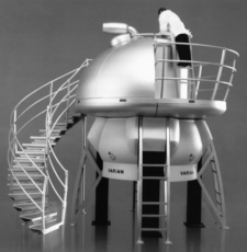
Figure 15. Equipment for high-resolution nuclear
a combination of two laser beams, one to illu-
magnetic resonance (NMR) spectroscopy.
minate and image the sample, and a second that
34
shapes the first beam and reduces the effects of
diffraction. The technique has been used to dis-
The amount of genetic data available
tinguish crystals only 100 nm apart and is still
and the rate of acquisition are astonish-
undergoing improvement.
ing by any measure.
Lasers, together with magnets, are being used
to develop technologies for manipulating single
The use of computers to model protein folding
molecules. Investigators are now able to exam-
is one of the primary efforts in the postsequenc-
ine how DNA interacts with the various protein
ing phase of the Human Genome Project. In the
molecules that cut, paste, and copy it. DNA is
1970s, when the first proteins were modeled,
an ideal choice for single-molecule studies. It
the structures generated were in vacuo (in a vac-
is a very large molecule (the longest human
uum), with no other molecules interacting with
chromosome stretches to 9 centimeters) and
the protein. Of course, each protein in a living
quite robust. For example, scientists have suc-
cell is surrounded by thousands of water mol-
ceeded in using lasers as optical tweezers to tie
ecules, and these have an important effect on
knots in single DNA molecules.2 Results indi-
the protein’s conformation. Indeed, research has
cate that knotted DNA is stronger than actin, a
demonstrated that the water-containing models
major muscle protein. Although tying DNA into
of proteins are much better predictors of how
knots may not seem particularly useful, it does
the proteins look and function within a cell.10
provide insight into the molecule’s mechanical
properties, which are critical to understanding
The importance of protein folding was recently
how enzymes interact with it.
recognized by IBM, which announced that it
would spend $100 million to build a supercom-
4.5 Simulations and computations
puter called Blue Gene. The five-year IBM initia-
The explosion of data produced by the Human
tive will involve modeling how proteins take on
Genome Project led to the creation of a new
their three-dimensional shapes. A major aim is
discipline, bioinformatics, whose focus is on
to help drug researchers identify drug targets for
the acquisition, storage, analysis, modeling, and
treating diseases. Protein folding is a daunting
distribution of the many types of information
problem. Even Blue Gene, which will be 500
embedded in DNA and protein-sequence data.14
times faster than the current fastest computer,
Biologists are familiar with the terms in vivo and
will require about one year to simulate the com-
in vitro, used to describe processes that occur
plete folding of a typical protein. The stakes,
in the body and in the test tube, respectively.
however, are huge. Approximately one-third
Now they are becoming acquainted with a new
of the genes identified in the newly sequenced
term, in silico, used to describe a new branch
human genome are of unknown function and
of biology that requires little more than a com-
are therefore of particular academic and com-
puter and a connection to the Internet. As more
mercial interest. New companies are formed
and more DNA and protein sequence data find
on a monthly basis to take part in this genetics
their way into computer databases, the ability
sweepstakes.
of bioinformatics to address biological ques-
tions becomes more powerful. The amount of
5 Technology and the Origins of
genetic data available and the rate of acquisition
Molecular Biology
are astonishing by any measure. According to
This section provides a brief history of the ori-
Francis Collins, head of the National Human
gins of molecular biology. It addresses the gene’s
Genome Research Institute, it took four years
chemical nature, organization, and behavior.
to obtain the first 1 billion base pairs of human
Despite molecular biology’s narrow focus on
sequence and just four months to get the second
DNA, it is readily apparent that many of the
billion.16
most important advances in the field have relied
35
Information about Using Technology to Study Cellular and Molecular Biology Using Technology to Study Cellular and Molecular Biology
heavily on technology-based contributions from
unaware of the important one-gene–one-enzyme
chemistry and physics. This is addressed in the
work of George Beadle and Edward Tatum from
National Science Education Standards. The His-
the early 1940s), the book has been credited
tory and Nature of Science Content Standard
with influencing a generation of physicists to
G states, “As a result of activities in grades 9 to
consider biological questions.
12, all students should develop understanding
of . . . historical perspectives.” It further states,
Soon, the ranks of the Phage Group began to
“Occasionally, there are advances in science and
grow. It included other physicists, such as Leo
technology that have important and long-lasting
Szilard, holder of the patent for the nuclear
effects on science and society.”
chain reaction and a participant in the Man-
hattan Project, and Thomas Anderson, one
Science historians often attribute the origins of
of the first American electron microscopists.
molecular biology to the Phage Group, which
Micrographs obtained by Anderson and Roger
first met in 1940 at Cold Spring Harbor Labora-
Herriott showed that phage begin the infection
tory in Long Island, N.Y. At the center of the
process by attaching to bacteria by their tails.
group were three scientists. Max Delbrück, a
Later, empty phage “ghosts” could be seen on
German physicist working at Vanderbilt Uni-
the bacterial surface.
versity, and Salvador Luria, an Italian biologist
working at Indiana University, had fled to the
Hershey and his colleague Martha Chase used
United States from Nazi Europe. They were
phage to examine the molecular nature of the
joined at Cold Spring Harbor by Alfred Hershey,
gene.11 They took advantage of radioactive iso-
an American biologist working for the Carnegie
topes that became available as a consequence
Institution’s Department of Genetics.
of work on the atomic bomb. Despite the ear-
lier work of Oswald Avery and his colleagues
Bacteriophage, also called phage, are viruses
demonstrating that DNA was the hereditary
that infect bacteria.1 These were discovered in
substance,3 many scientists continued to believe
1916 by the English microbiologist F.W. Twort
that genes could only be made of protein. Her-
and, independently, two years later by the
shey and Chase began their experiment by
French-Canadian F. d’Herelle. It was d’Herelle
using radioactive phosphorous to label phage
who came up with the name bacteriophage.
DNA and radioactive sulfur to label phage pro-
Phage became an important area of research in
tein. They tried to detect which radiolabel went
the 1920s, when scientists hoped they could be
inside the bacterium to direct synthesis of new
used to treat bacterial diseases. When this hope
phage particles after the bacterium was infected.
failed to materialize, phage research fell out of
At first, they could not effectively detach the
favor until the Phage Group resurrected it.22
phage particles from the surfaces of the bacte-
rial cells, but then an unexpected technology
In 1944, Delbrück organized a summer course
came to their aid. They used a Waring blender,
at Cold Spring Harbor Laboratory to introduce
originally designed to mix cocktails, to disrupt
other scientists to the quantitative methods for
the attachments of the phage to the bacterial
studying phage that he and Luria had devel-
cells. The radioactive phosphorous went into
oped. In that same year, the great Austrian
the bacterial cells, while the radioactive sulfur
physicist Erwin Schrödinger published a book
remained outside with the phage ghosts, con-
titled What Is Life? that discussed heredity from
firming that DNA, and not protein, contains the
a physics perspective.19 Schrödinger reasoned
genetic information. This work set the stage for
that although living things obey the laws of
the contribution of the youngest member of the
physics, they also might be governed by undis-
Phage Group, James Watson.
covered physical laws. Although biologists of
that time regarded Schrödinger’s book as roman-
Watson came to the Cavendish Laboratory at
tic and a bit naive (for example, he seemed
Cambridge University in 1951, ostensibly to
36
study the three-dimensional structures of pro-
Samples of cells were removed before the switch
teins. He quickly fell in with Francis Crick, a
to the light-isotope growth medium (genera-
British physicist, who had developed an inter-
tion 1) and from the first two generations fol-
est in heredity after reading Schrödinger’s What
lowing the switch (generations 2 and 3). DNA
Is Life? The pair formed a collaboration that
samples extracted from the cell samples were
resulted two years later in the proposal of the
centrifuged through a solution of cesium chlo-
double helix model of DNA.23 Although Watson
ride that forms a density gradient during cen-
and Crick relied on model building to solve
trifugation (for 20 hours at 40,000 revolutions
DNA’s structure, they could not have succeeded
per minute). DNA molecules form a discrete
without help from two other scientists at Cam-
band at a position where their density equals
bridge, Maurice Wilkins and Rosalind Franklin.
that of the cesium chloride gradient. The DNA
Wilkins first, and then Franklin, used X-ray
samples taken from generation 1 contained a
diffraction to study the structure of DNA. In
single heavy band, since both DNA strands con-
the case of DNA fibers, the diffraction patterns
tained the 15N isotope. Samples from generation
suggested that the molecule was some type of a
2 displayed a single band of medium density,
helix with a diameter of 20 Å and a repeat of 34
since each DNA molecule consisted of one
Å. Near the end of the paper that describes the
heavy (15N) parental strand and one light (14N)
double helix, Watson and Crick included the
complementary strand. Finally, samples from
statement, “It has not escaped our notice that
generation 3 displayed bands of two different
the specific pairing we have postulated immedi-
densities. One band of medium density again
ately suggests a possible copying mechanism for
consisted of a heavy parental strand and a new
the genetic material.”
complementary light strand. A second band of
light density consisted of two strands of light
Experimental support for a copying mechanism
DNA, one an inherited light parental strand and
suggested by the double helix structure came in
the other, a new complementary light strand.
1958 from Matthew Meselson and Frank Stahl,
then working at the California Institute of Tech-
Around the time that Meselson and Stahl were
nology. In what some have called “the most
performing their experiments, Crick theorized
elegant experiment in molecular biology,”