CLASSIFICATION OF GENETIC DISORDERS:
1. Chromosomal Disorders
2. Monogenetic conditions (Single gene disorder)
3. Polygenetic conditions (Multifactorial disorder)
4. Disorder with non-traditional mechanism of expression and inheritance
1. Chromosomal disorders:
These can be due to abnormality in number or structure of genes such as:
Rearrangement of chromosomal disorders can be further divided into:
2. Monogenetic conditions:
These follow Mendelian law of inheritance. Monogenetic conditions can be divided into four mode of inheritance:
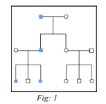
FIG 1: A pedigree illustrating an autosomal dominant disorder. Affected individuals are depicted by colour. By convention, males are represented by square and females by circle.

FIG 2: A pedigree illustrating an autosomal recessive disorder. Affected individuals are depicted by solid colour and heterozygotes by partial shading.
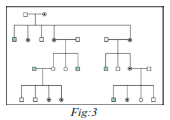
FIG:3 A pedigree illustrating an X linked recessive disorder. Affected individuals are depicted by solid colour and heterozygous carrier by dots.
3. Polygenetic Conditions:
These are multifactorial disorders, determined by nongenetic or environmental factors. Polygenetic conditions do not follow Mendelian law of inheritance.
4. Non-traditional Mechanism of Expression & Inheritance:
Non-traditional mechanism of expression and inheritance occurs due to mutation in mitochondrial genome. It can be due to uniparental disomy i.e. two alleles from same locus inherited from one parent instead from both the parents. Hence it is also known as parent-of-origin effect.
MITOCHONDRIAL INHERITANCE:
Majority of genes are enclosed inside the nuclear genome, however there are small number of gene located on mitochondrial DNA (mt DNA). The mtDNA consist of 16.kb of circular DNA and accounts for 1% of total DNA. It encodes protein for aerobic respiration. Each cell having functional mtDNA will be identical (homoplasmy). However mutation in mtDNA will lead to a mix blend of expression (heteroplasmy). The proportion of mitochondrial mutated DNA will determine the phenotypic expression of mutation. More than 85% of mtDNA mutation before there is a phonotypical significant defect in aerobic respiratory chain.
To diagnose the disorders due to mutation in mtDNA muscle biopsy is profound over blood sampling to rule out false report, as in mtDNA mutation disorder there may be variation among different body tissues.
MUTATION: Mutation can be defined as alternation or change in genomic material. Mutation can be of two types depending upon the cell line involved:
At molecular level structural mutation can be majorly divided into following types:
1. Single base pair substitution: It involves substitution of one base pair for anther. Single base pair mutation is also known as point mutation. They can further be divided into :
2. Deletion: These may vary in size from single base pair to mega base pair. E.g. More than 60% of mutations in Duchenne muscular dystrophy are due to deletion in dystrophic gene.
3. Insertion & Duplication: They occur as result of unequal crossing over at meiosis.
4. Frameshift mutation: They can occur either due to deletion or single base pair substitution resulting in misreading of subsequent following codon.
5. Dynamic mutation: These occur when triplet of nucleotide repeated in a gene and the transmission of exact number of repeat is unstable and may increase in further generation. E.g. Fragile X syndrome, Mental retardation.
6. Inversion: It occurs when there is break in two chromosomes, the segments flip over and re-join, resulting in in correct translation.