Skin rash
Test 2
+ for poison
Poison ivy
Steroid skin
ivy
cream
16
G
Sore throat
Test 1
+ for bacteria
Strep throat
Antibiotic
A
17
G
Sore throat
Test 1
+ for bacteria
Strep throat
Antibiotic
A
18
B
Ankle pain
No test
X-ray
No break
Bandage
19
G
Sore throat
Test 1
+ for bacteria
Strep throat
Antibiotic
A
20
D
Sore toe
Test 1
– for all
Blister from Dressing
bacteria
boots
21
C
Sore toe
Test 1
+ for bacteria
Blister from Dressing
A
boots
Copyright © 2011 BSCS.
Master 2.3
Permission granted for classroom use.
Follow-up on Week 1 Infi rmary Visits
Summary of Patients’ Responses to Treatment from Week 1
• Six soldiers from Barracks A with pinkeye tested positive for infection by bacterial species C. They were treated with eyedrops containing antibiotics.
In all cases, the infection has cleared up.
• Six soldiers from Barracks G with sore throats tested positive for infection by bacterial species A. They were treated with antibiotics. Although two soldiers returned to the infi rmary, all are responding to the antibiotics and the infections have largely disappeared.
• Four soldiers developed blisters from wearing new boots. Three of the four tested positive for infection by bacterial species A, and the fourth soldier tested negative for species A, B, and C. One soldier received a cut on the left leg, which was closed with stitches. He has no evidence of infection.
• One soldier twisted his ankle. X-rays showed no broken bones. The ankle was bandaged, and the soldier has been assigned to light duty.
• Two soldiers from Barracks E tested positive for exposure to poison ivy. They were treated with steroid cream, and the skin rashes are disappearing.
• One soldier, who was short of breath, was diagnosed as having asthma and was given an inhaler, which eased her symptoms.
Copyright © 2011 BSCS.
Master 2.4
Permission granted for classroom use.
Visits to the Infi rmary, Week 2
Return Visits to the Infirmary from Week 1 Soldier Visits
Soldier Barracks What’s wrong? Tests ordered Test results Diagnosis Treatment 2
D
Swollen,
blistered
lower leg
16
G
Sore throat
19
G
Sore throat
New Visits to the Infirmary, Week 2
Soldier Barracks What’s wrong? Tests ordered Test results Diagnosis Treatment 22
G
Sore throat
23
A
Sore throat
24
E
Cut on head
25
G
Sore throat
26
G
Sore throat
27
B
Skin rash
28
G
Sore throat
29
D
Ankle pain
30
A
Sore throat
31
B
Sore toe
32
F
Skin rash
Copyright © 2011 BSCS.
Master 2.5
Permission granted for classroom use.
Medical Reference Manual:
Necrotizing Fasciitis (Flesh-Eating Bacteria)
What is it?
Flesh-eating disease is a bacterial infection that destroys skin and fat tissue. The disease is very rare. The odds of getting it are about 1 in 100,000. However, it is very serious. About 2 out of 10 people who get this infection die from it.
What causes it?
The disease can be caused by different species of bacteria, including the one that causes strep throat. The bacteria enter the body through open wounds, where they interact with the immune system to produce the disease. Flesh-eating disease is rare because the immune system of most people will stop the infection before it becomes serious.
What are the symptoms?
The skin reddens, becomes swollen, and is painful to the touch. Other symptoms include nausea, vomiting, and diarrhea. The symptoms start suddenly, may get better for a day or two, then quickly worsen. If not treated, the disease may result in organ failure and death.
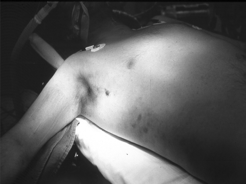
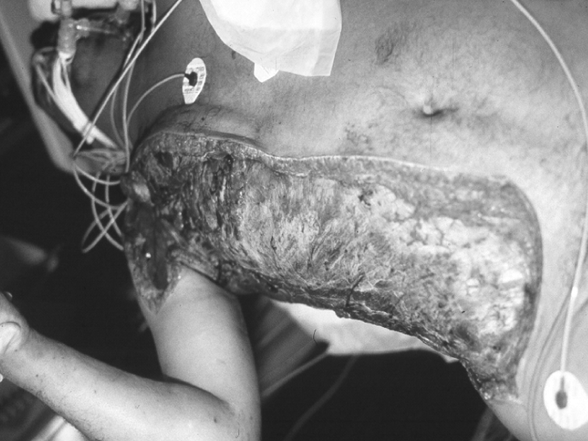
Figure 1. Early infection. (Donald E. Low, University
Figure 2. Late infection. (Donald E. Low,
Health Network/Mount Sinai Hospital)
University Health Network/Mount Sinai Hospital)
How is it treated?
Patients with flesh-eating disease need immediate hospital care. Treatment involves antibiotics and surgery to remove diseased tissue and stop the spread of the disease.
Copyright © 2011 BSCS.
Master 2.6
Permission granted for classroom use.
Questions about a Rare Disease
Name(s):
1. What evidence suggests that bacterial species A causes both sore throat and foot infection?
2. What evidence suggests that the soldier with the foot infection has fl esh-eating disease?
3. Why are there many cases of sore throat but only one case of fl esh-eating disease?
4. What evidence is there that fl esh-eating disease is a rare disease?
5. What should be the next step in treating the soldier with the foot infection?
Explain your reasoning.
Copyright © 2011 BSCS.
Master 2.7
Permission granted for classroom use.
To Play or Not to Play?
Patrick is a 13-year-old middle school student who loves to play basketball. He came home excited from school and explained to his parents that the school basketball team will be holding tryouts next month and he wants to participate.
Patrick’s parents are both happy and concerned for him. They are happy because they know Patrick loves sports, and they feel that the exercise will be good for him. They also know that Patrick has been occasionally teased because he is tall and thin. Maybe by joining the basketball team he will make new friends and feel more accepted by his classmates.
Patrick’s parents are concerned because he has some health problems. When Patrick was a toddler, the family doctor diagnosed him with a heart murmur.
The doctor explained that a heart murmur refers to a sound that the blood makes as it flows through the heart. She further explained that heart murmurs are usually harmless and that Patrick could lead a normal life.
When he was nine, Patrick developed a problem with his eyesight, and it was discovered that one of his eye lenses was detached and had to be repaired.
When Patrick was 10, he was diagnosed with asthma. The doctor explained that asthma causes the tubes carrying air in and out of the lungs to become sore and swollen. This can cause coughing and wheezing and make it difficult to breathe.
The doctor created a treatment plan for Patrick that helped him recognize his symptoms and use an inhaler to make breathing easier. She also explained that, with proper management of his asthma, Patrick could play sports and that the exercise might even improve his condition.
Finally, just last year, Patrick was diagnosed with scoliosis, or curvature of the spine. The doctor explained that Patrick’s scoliosis was moderate and, as with most children, the cause was unknown. He further explained that in 90 percent of cases, no future treatment is needed.
Copyright © 2011 BSCS.
Master 3.1
Permission granted for classroom use.
Medical Specialty Report Form
Name(s):
Patient’s name
Medical specialty
Patient’s medical history
Results from physical exam
Possible causes
Copyright © 2011 BSCS.
Master 3.2
Permission granted for classroom use.
Heart and Circulatory System
Cardiologist Report
Medical history
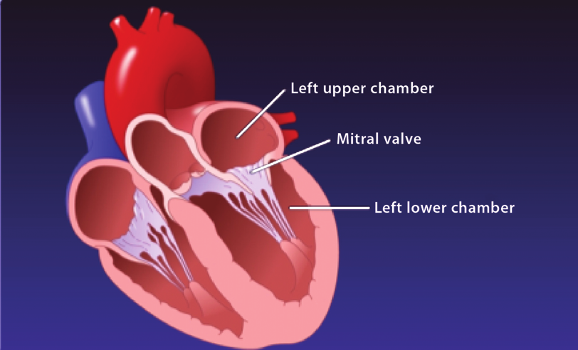
Patient was previously diagnosed with a heart murmur. An echocardiogram reveals mitral valve prolapse and an enlarged aorta.
Physical exam
The presence of a heart murmur was confirmed. An echocardiogram revealed the presence of mitral valve prolapse.
Copyright © 2011 BSCS.
Master 3.3
Permission granted for classroom use.
(Page 1 of 2)
Medical Reference Manual: Heart and Circulatory System
Heart murmur
When doctors use a stethoscope to listen to the heartbeat, they hear a lub-DUB sound made by heart valves opening and closing as blood flows through the heart. The term
“heart murmur” refers to an unusual whooshing sound doctors hear when listening to the heartbeat.
Doctors diagnosis heart murmurs in many children at some point in their lives. Most heart murmurs are harmless and need no treatment. Other heart murmurs are called abnormal and may be associated with defects in the heart that were present at birth.
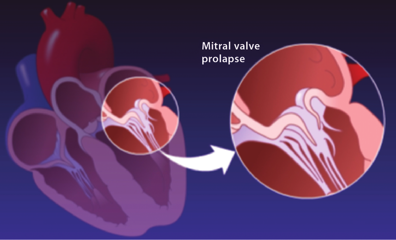
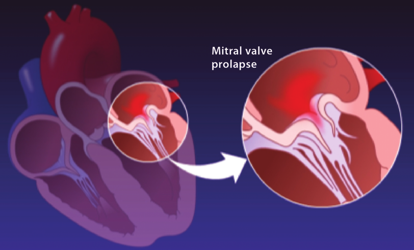
Mitral valve prolapse
In the condition called mitral valve prolapse, one of the heart’s valves doesn’t work properly. The valve flaps are “floppy” and don’t close properly. This sometimes causes blood to flow backward from its normal direction. This backflow of blood may be associated with shortness of breath or chest pain.
The cause of mitral valve prolapse is not known. Most people with the condition are born with it. It tends to run in families and is associated with connective tissue disorders such as Marfan syndrome.
Figure 1. Mitral valve prolapse.
The mitral valve
is a valve that lies
between the left
upper and lower
chambers of the
heart.
In mitral valve prolapse, the valve
This lack of a tight seal can cause
flaps are too large and don’t form a
a small amount of blood to flow
tight seal when they close.
backward, resulting in a heart murmur.
Copyright © 2011 BSCS.
Master 3.3
Permission granted for classroom use.
(Page 2 of 2)
Vision System
Ophthalmologist Report
Medical history
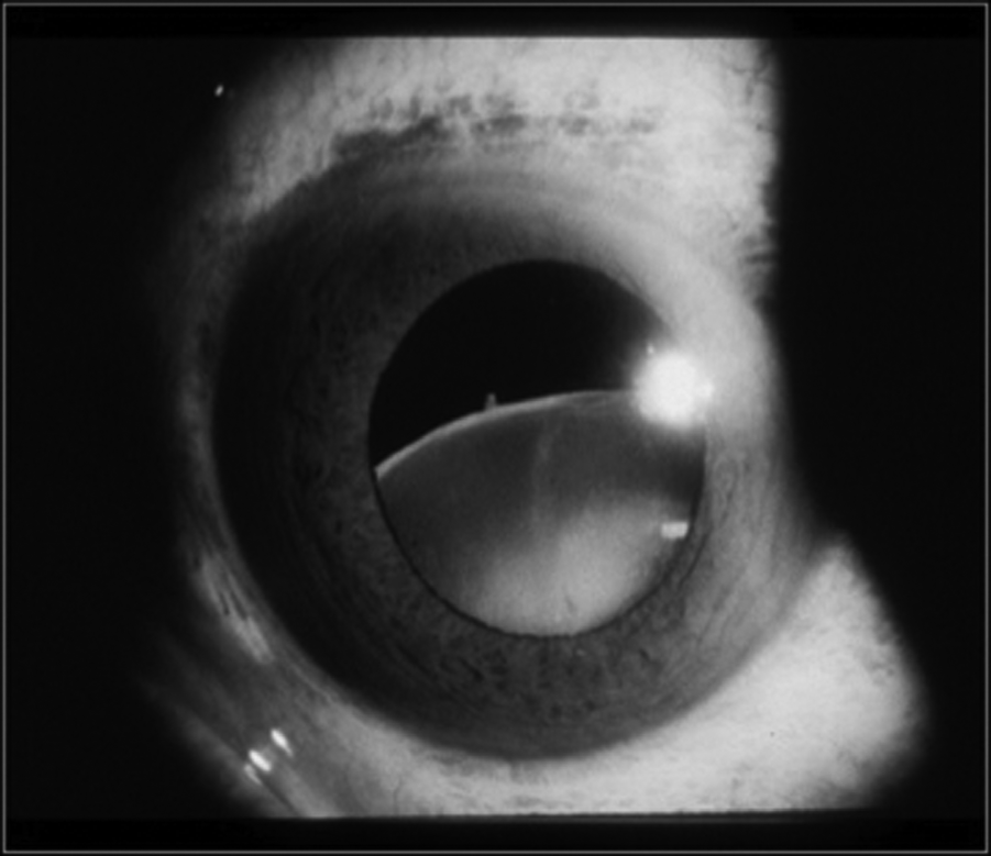
The patient is myopic (nearsighted). When he was nine years old, he was being fitted for eyeglasses when an exam revealed that his left lens was dislocated.
Physical exam
An eye exam confirmed myopia and a repaired detached left lens.
Figure 1. Slit lamp exam: Photo from a slit lamp exam of a patient’s eye showing a detached left lens. (Kevin J. Blinder, MD, The Retina Institute, Washington University School of Medicine)
Copyright © 2011 BSCS.
Master 3.4
Permission granted for classroom use.
(Page 1 of 2)
Medical Reference Manual: Vision System
Myopia (nearsightedness)
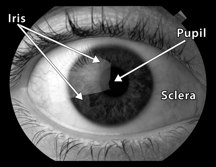
Nearsightedness is caused by a change in the shape of the eyeball so that it is egg shaped instead of round. This egg-shaped eyeball focuses light a little in front of the retina instead of directly on it, resulting in blurry vision. Myopia is a common condition affecting 30 to 40 percent of the American population.
Detached lens
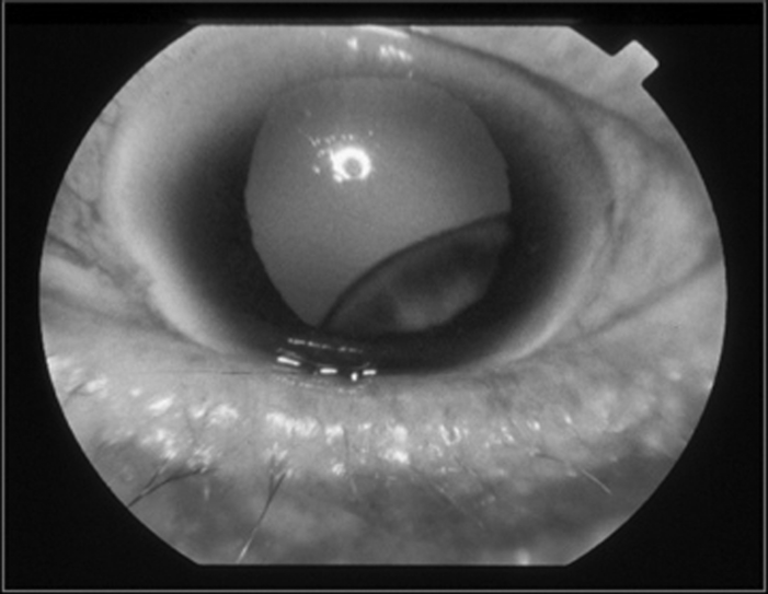
A detached, or dislocated, lens means that the lens has shifted from its normal position (centered behind the pupil). If the dislocation is moderate, the problem may be corrected with glasses. A severe lens dislocation may require surgery to correct.
Detached lenses are rare in the general population. They are often caused by a blow to the eye. The condition is much more common among people with certain diseases involving connective tissue such as Ehlers-Danlos syndrome and Marfan syndrome.
Figure 2. Photos from slit lamp exams: Left, a normal lens; right, a detached lens.
((left) Corbis, (right) Kevin J. Blinder, MD, The Retina Institute, Washington University School of Medicine) Copyright © 2011 BSCS.
Master 3.4
Permission granted for classroom use.
(Page 2 of 2)

Respiratory System
Pulmonologist Report
Medical history
Patient has been diagnosed with asthma. When seven years old, he experienced a collapsed lung.
Physical exam
Exam confirmed the diagnosis of asthma. Lung volume was normal. Chest X-ray was normal.
Figure 1. Patient photo: Patrick at age 10.
Copyright © 2011 BSCS.
Master 3.5
Permission granted for classroom use.
(Page 1 of 2)
Medical Reference Manual: Respiratory System
Table 1. Information about Asthma
What are the
• Coughing
symptoms of
• Wheezing
asthma?
• Diffi culty breathing
What causes asthma Asthma attacks are a response to environmental triggers attacks?
that leads to
• Infl ammation of the airways. This swelling and irritation
inside the airways leads to diffi culty breathing.
• Bronchospasms. The muscles surrounding the airways
go into spasm, leading to their narrowing.
In rare cases, asthma is associated with disorders of the
connective tissue.
What are the
• Dust
triggers for asthma
• Mold
attacks?
• Pollen
• Pets
• Cigarette smoke
• Pollution
• Illness (infection by bacteria and viruses)
How is asthma
Inhaled drugs, such as albuterol, help widen the airways
treated?
during asthma attacks and make it easier to breathe.
For long-term management of asthma, inhaled steroids are
safe and can be used every day.
Copyright © 2011 BSCS.
Master 3.5
Permission granted for classroom use.
(Page 2 of 2)
Skeletal System
Orthopedist Report
Medical history
Patient was previously diagnosed with mild scoliosis (curvature of the spine).
Physical exam
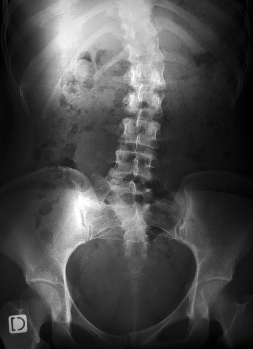
A physical exam and X-rays confirmed the presence of mild scoliosis. The curvature was measured to 15 degrees. It was noted that the patient has unusually long, slender arms, fingers, and feet.
Figure 1. Patient X-ray: X-ray taken when patient was fi ve years old.
(© Ldambies | Dreamstime.com)
Copyright © 2011 BSCS.
Master 3.6
Permission granted for classroom use.
(Page 1 of 2)
Medical Reference Manual: Skeletal System
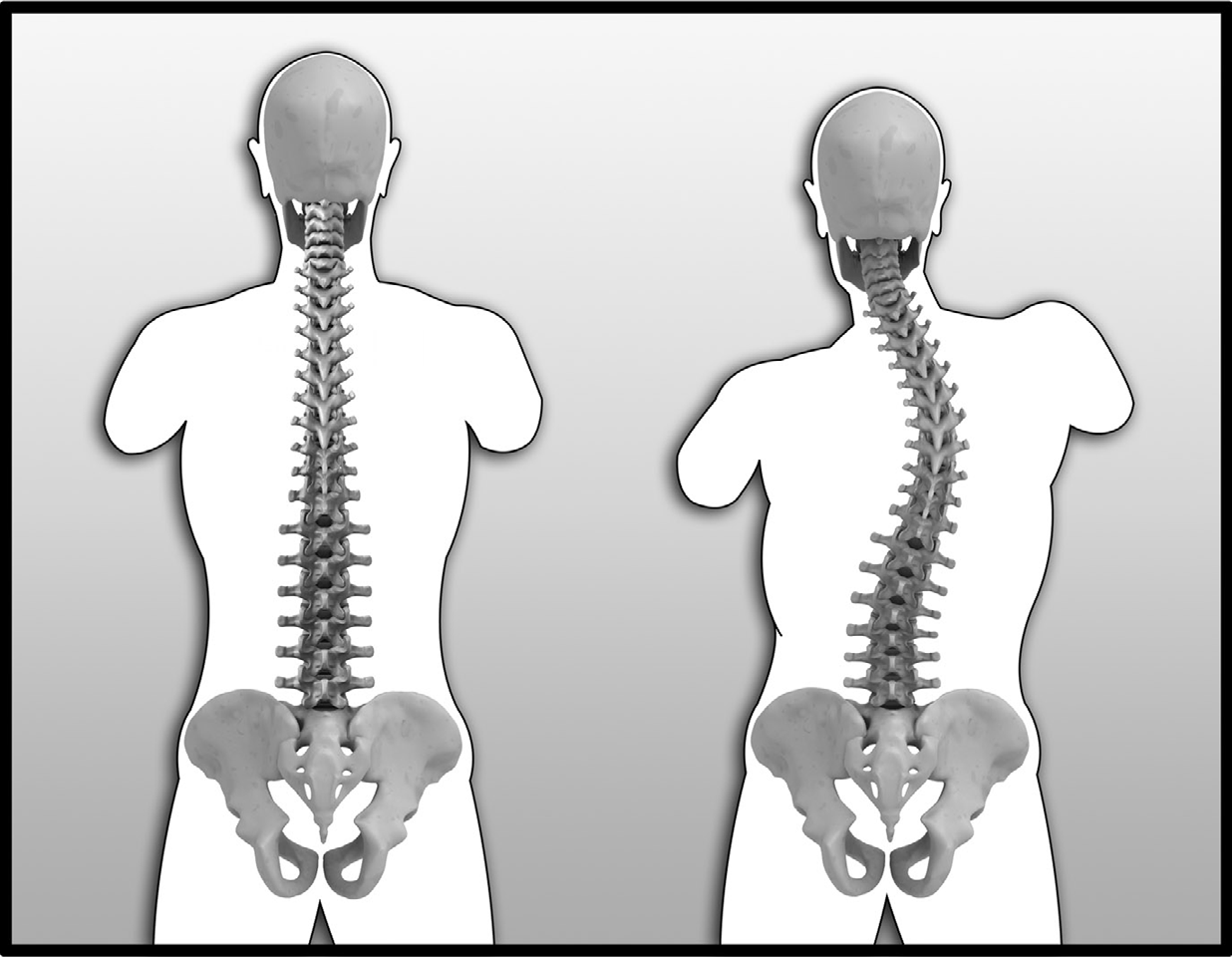
Scoliosis (curvature of the spine)
Scoliosis refers to an abnormal curvature of the spine. About 10 percent of adolescents show some degree of scoliosis, but less than 1 percent need treatment for the condition. The severity of scoliosis is described by the extent of the curvature.
Table 1. Severity of Scoliosis
Amount of Curvature
Severity
Curvature less than 20 degrees
Mild
Curvature between 20 and 70 degrees Moderate
Curvature greater than 70 degrees
Severe
Patients with mild scoliosis usually don’t require treatment beyond examination to see whether the condition worsens. Patients with moderate and severe scoliosis are treated with back braces or surgery.
In most cases, the cause of scoliosis is not known; however, it does seem to run in families. In some cases, the condition is caused by an injury. In other cases, the condition is a result of a muscle, nerve, or connective tissue disease.
Figure 2. The spine and scoliosis: Left, normal spine; right, spine showing scoliosis.
(© Sebastian Kaulitzki | Dreamstime.com)
Copyright © 2011 BSCS.
Master 3.6
Permission granted for classroom use.
(Page 2 of 2)
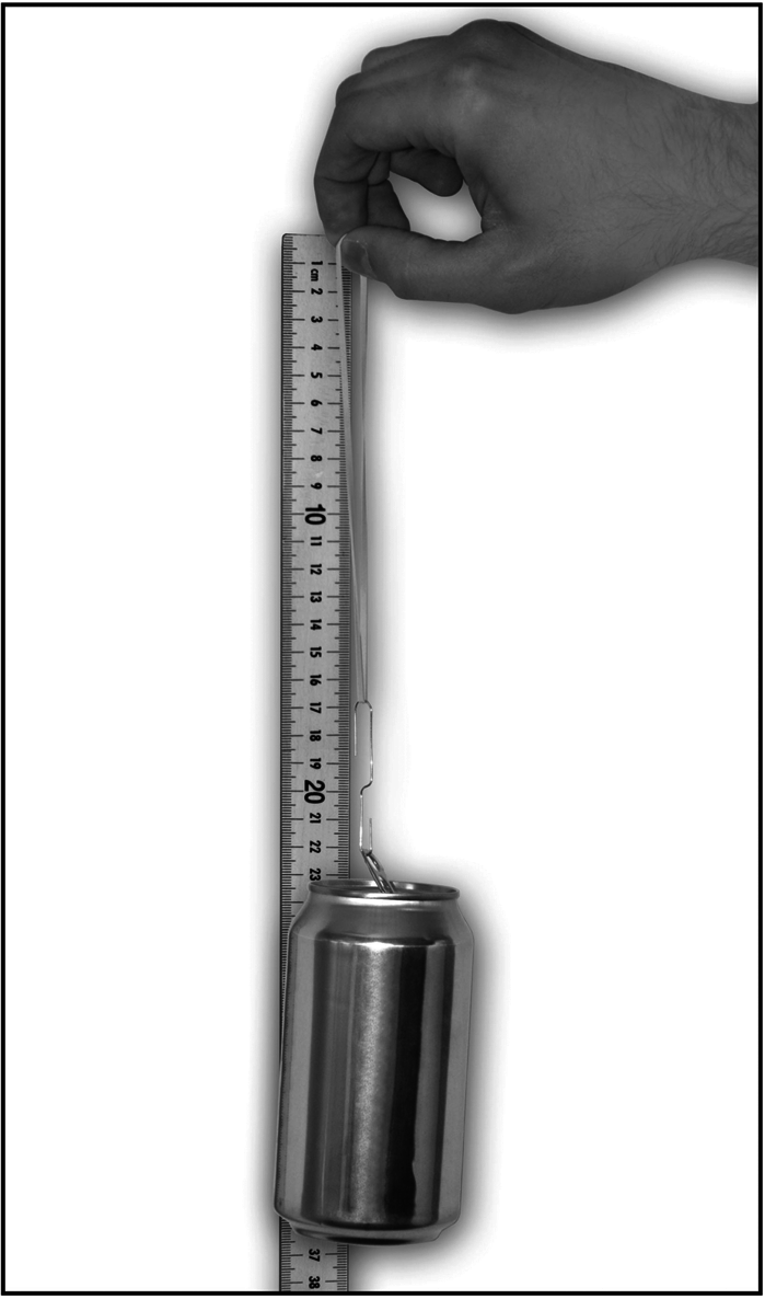
Measuring Elasticity
Steps for measuring the elasticity of rubber bands
1. Unfold the paper clip into an S shape.
Figure 1. Measuring elasticity.
2. Slip one end of the paper clip
through the pulled tab on top of the
soda can.
3. Place the other end of the paper clip
through the rubber band.
4. Hold a meter stick upright on a hard
surface so that the end reading “100
centimeters” is resting on the surface
and the end reading “0 centimeters”
is up in the air.
5. Hold the top of rubber band (the end
away from the paper clip) up to the
end of the meter stick that reads “0.”
6. Observe and record in your notebook
how far down the meter stick the
rubber band has stretched.
7. Repeat Steps 2 through 6 for the
second rubber band.
Copyright © 2011 BSCS.
Master 3.7
Permission granted for classroom use.
Diagnosing a Connective Tissue Disorder
Name(s):
1. You will try to match Patrick’s medical symptoms to four different disorders of connective tissue.
2. For each of Patrick’s symptoms listed in the table under “medical history,”
decide whether that symptom is consistent with each of the four connective tissue disorders written across the top row. Use the information supplied in the Medical Reference Manual.
3. Place a check mark in the appropriate box when the symptom is consistent with the connective tissue disorder.
Table 1. Checklist of Patrick’s Symptoms
Patrick’s
Ehlers-Danlos
Marfan
Osteogenesis
Scleroderma
medical history
syndrome
syndrome
imperfecta
Myopia
Detached eye
lens
Asthma
Collapsed lung
Heart murmur
Leaky heart valve
Long arms and
legs
Curvature of
spine
Copyright © 2011 BSCS.
Master 3.8
Permission granted for classroom use.
Medical Reference Manual: Disorders of the Connective Tissue
Connective tissues are made of proteins and fats. They support your body’s organs and give your tissues their shape. Cartilage is an important connective tissue. It is stiff but more flexible than bone. Cartilage helps your bones move and glide over each other.
It also gives shape to body parts such as your nose and ears.
Connective tissue may be damaged by injury or through an infection. It can also be damaged by a large number of genetic disorders that occur rarely in the population.
A few of them are described below.
Ehlers-Danlos syndrome
Ehlers-Danlos syndrome refers to a collection of related disorders that weaken connective tissues. Symptoms can be mild to life threatening. They include the following:
• heart valves that leak
• weakened blood vessels
• loose joints
• abnormal wound healing
• soft, stretchy skin that bruises easily
• muscle weakness
• joint dislocations
Ehlers-Danlos syndrome is an inherited disorder. Treatment involves managing symptoms and learning how to protect the joints and prevent injuries.
Scleroderma
Scleroderma is a group of related disorders involving abnormal growth of connective tissue. One type of scleroderma affects only the skin. Another type can also affect other body systems. The cause of scleroderma is not known. It is more common in females than males. Other symptoms may include the following:
• calcium deposits in connective tissues
• narrowing of blood vessels in the hands and feet
• swelling of the esophagus (tube between the throat and stomach)
• thick, tight skin on fi ngers
• red spots on hands and face
Treatment involves managing the symptoms.
Copyright © 2011 BSCS.
Master 3.9
Permission granted for classroom use.
(Page 1 of 2)
Marfan syndrome
Marfan syndrome is a disorder of connective tissue that is due to mutations in a gene that codes for a connective tissue protein called fibrillin. Symptoms can be mild to severe. Often, people with Marfan syndrome are tall and thin and have loose joints.
Their fingers and feet may be unusually long. Other symptoms may include the following:
• heart valves that leak
• heart murmur
• weakened blood vessels
• curvature of the spine
• fl at feet
• sudden lung collapse, sometimes asthma
• nearsightedness and problems with the eye lens
• stretc