the scope of functions
5x + 14 = 34. What is the value of x?)
regulated by the
• recall and describe the way to get from the classroom to the
cafeteria (e.g., Give directions to walk from this classroom
human brain.
to the cafeteria.)
• read a sentence aloud (e.g., Read the following sentence aloud:
“Four score and seven years ago our fathers brought forth on this
continent, a new nation, conceived in Liberty, and dedicated to
the proposition that all men are created equal.”8)
2. After the volunteers perform the tasks, ask the students to identify
the part of the body that is involved in all of the tasks.
The goal for this question is for students to acknowledge that the
brain is involved in regulating all human physiological, behavioral,
and emotional functions. For example, point out that all students
are breathing. When most people think about breathing, they think
about the lungs, but not the involvement of the brain. Also, point out
that each student’s heart is beat ing. Although the heart is actually
pumping the blood, the brain fulfills an important role in regulating
the heartbeat. The involvement of the brain will be more obvious for
some of the tasks than for others.
3. After students deduce that the brain is involved in all of these
activities, ask students to suggest how they think scientists
investigate what hap pens in the human brain.
Students will provide a variety of answers, including watching a
person’s behavior, using various imaging techniques (such as PET
scans, CT scans, or MRI), using animals (either living or dead) for
research, and so forth.
28
Activity 2: Positron Emission Tomography and
Brain Function
The following procedure describes how to conduct the
Web version of this activity, which is the preferred method
Content Standard A:
of instruction. Instructions for conducting the alternative
Formulate and revise
print version follow the shaded Steps 1–5.
sci entific explanations
and models using logic
1. Before starting the Web-based activity, inform students that they
and evidence.
will be analyzing positron emission tomography (PET) images.
Content Standard A:
PET is one technology scientists use to learn about the function
of the living human brain. The PET images that the students will
Scientists rely on
examine use radioactive glucose to identify parts of the brain that
technol ogy to enhance
are active. Active brain areas use more glucose than less active areas
the gath ering and
and thus more of the labeled glucose is taken up into the active areas.
manipulation of data.
PET images are color-coded by a computer. The most active brain
Content Standard C:
areas are shown in red. Areas in yellow are less active than areas
Organisms have
in red, but are more active than areas in green. The least active areas
behav ioral responses to
are shown in blue or purple. Stu dents will see a color scale on the
screen with the PET images for reference.
inter nal changes and to
external stimuli.
Students may have seen color-coded computer images on television
weather reports. In weather radar images, areas encountering heavy
storms appear in red and yellow, and areas experiencing milder
weather distur bances appear in green or blue.
2. Divide the class into groups of three students. Arrange for each
group to work at a computer to complete the online activity
Analyzing Brain Images. Give each group a copy of Master 1.2,
Go to the supplement’s Web site . Select Lesson 1— The Brain: What’s Going On in There?
3. Instruct students to work with their group members to analyze
the PET images and to answer the questions on Master 1.2.
When students reach question 5, display a transparency of
Master 1.3, PET Image Tasks, to provide the needed information.
4. After the groups complete the activity and write their answers
to the questions on Master 1.2, discuss the answers to the
questions as a class.
29
Student Lesson 1
The Brain: Understanding Neurobiology Through the Study of Addiction
Sample Answers to Questions on Master 1.2
Question 1. When you look at the images that make up Set #1
(Master 1.1), how do the four images differ from each other?
Content Standard A:
Communicate and defend
The brain images are different sizes. The images show variation in
a scientific argument.
the amount and pattern of the different colors.
Question 2. Why are four images shown in each set of PET images?
Why would scientists need to examine more than one PET image
taken of a sub ject’s brain?
The four PET images in each set show the activity at different levels
of the brain. If a scientist examines only a single image, he or she
increases the chances of missing important information.
Question 3. When comparing the images in Set #1 with the images in
Sets #2, 3, 4, 5, and 6, how is the activity of the brain in each of
these sets different from Set #1’s?
Identify the image
that shows the
Set
greatest change
Number
(a, b, c, or d)
Describe the change in brain activity
2
b
There is more red on the right side of the
brain, mainly near the center in terms of
front-to-back direction. There is also red
on the left side, but it is not as strong as
it is on the right side.
3
b
The main activation is in the back of the
brain on both sides of the midline.
4
c
The main activation is at the front of the
brain near the periphery on both sides of
the midline.
5
d
The main activation is in four areas, two
on each side of the brain. Two are very
near the back of the brain, and two are
farther forward.
6
a
The main areas of activation are a spot
on the left side of the brain and a
smaller spot near the front of the
brain on the midline.
Question 4. The PET images shown in Set #1 show brain activity in
a rest ing brain. The images in Sets #2–6 show activity in the brains
of humans who are doing different tasks. When you look at the PET
scans and the chart in question #3, what generalizations can you make
about the activ ity of the brain when different tasks are performed?
30
The key points of this exercise are that different brain areas are
activated during dif ferent tasks and different brain functions are
localized to different brain areas.
Question 5. Compare the tasks that the subject performed during
each of the PET scans (as shown on the overhead transparency) with
the individual’s brain activity. Use the information from the overhead
and from the PET images to complete the following chart (Master 1.2b).
Set
Brain region that is more
This region is involved in
Number
active in the PET image
processing information related to
2
auditory cortex
hearing
3
primary visual cortex
vision, sight
4
frontal cortex
thinking
5
hippocampus
memory
6
motor cortex
movement
5. Instruct students to watch the online video How PET Works.
From the main menu, select Lesson 1— The Brain: What’s Going On in
There? Then click on How Is PET Done? This video expands students’
understanding of PET. A scientist explains how PET imaging is done.
Content Standard E:
Science often advances
After students have completed the activity, you may wish to challenge
with the introduction of
them by asking them to propose an explanation for why functions are
new technology.
local ized to specific brain areas. Why would this be beneficial from an
evolu tionary standpoint? (See Background Information on pages 20–21.)
The following procedure is for classes using the
print version of the activity.
1. Tell students that one of the ways that scientists investigate the
Content Standard A:
function of the living human brain is by using positron emission
Formulate and revise
tomography (PET). The PET images that the students will examine
scientific explanations
use radioactive glucose to identify parts of the brain that are active.
and models using logic
Active brain areas use more glucose than less active areas and thus
and evidence.
more of the labeled glu cose is taken up into the active areas. PET
scans are color-coded. The scale bar shown on Master 1.1, Positron
Content Standard A:
Emission Tomography (PET) Images, provides a reference. The most Scientists rely on
active brain areas are shown in red. Areas in yellow are less active
technology to enhance
than areas in red, but are more active than areas in green. The least
the gathering and
active areas are shown in blue or purple.
manipulation of data.
Content Standard C:
PET images are color-coded by computer to show activity in the brain.
Organisms have behavioral
This is similar to color-coded images students may have seen on
television weather reports. In weather radar images, areas encountering responses to internal
heavy storms appear in red and yellow, and areas experiencing milder
changes and to external
weather distur bances appear in green or blue.
stimuli.
31
Student Lesson 1
The Brain: Understanding Neurobiology Through the Study of Addiction
2. Divide the class into groups of three students. Give each group a
copy of Master 1.1, Positron Emission Tomography (PET) Images,
and a copy of Master 1.2, Interpreting PET Images.
3. Help students understand how the PET images correlate to the
orienta tion of the brain in the body.
The PET images show a cross-section of the brain. The four images in
each set show four different levels of the brain. In these images, the
front of the brain is toward the top (the subject’s face is toward the top
of the image). Have the students examine the PET scans and identify
the regions that become active in response to each stimulus.
4. Instruct students to work with their group members to answer the
ques tions on Master 1.2. When students reach question 5, display
a trans parency of Master 1.3, PET Image Tasks, to provide the Content Standard A:
needed information.
Communicate and defend 5. Discuss the answers to the questions on Master 1.2 as a class.
a scientific argument.
Sample answers for the questions on Master 1.2 are listed in the
procedure for the online version of Activity 2 on pages 30–31.
Activity 3: Parts of the Brain
Note to teachers: This activity is intended for classes that want more
Content Standard C:
informa tion about the anatomy of the brain. Learning the names and
Cells can differentiate
functions of brain lobes and regions is not a major focus and could distract
and complex multicellu lar
some students from the main concept, that brain functions are localized
organisms are formed
to specific brain areas. Understanding the main concept is critical for
understanding how neurons communicate and how drugs of abuse affect
as a highly organized
neuronal function. These topics are covered in Lessons 2 and 3.
arrangement of
differenti ated cells.
For classrooms using the Web version of this activity.
1. Have students continue in their groups to conduct the
online activity What Does This Part of the Brain Do?
To access this activity, go to the supplement’s Web site and select Lesson 1— The Brain: What’s Going On in There? Then click on What Does This
Part of the Brain Do?
2. Ask students to take out their completed worksheet on Master 1.2.
Review the tasks that the students performed in Step 1 of Activity 1
and ask students to identify the part of the brain that was active
in each case.
From the information in question 5 on Master 1.2, students should be
able to identify the brain area involved in some of the tasks performed
in Step 1, but others were not covered in that question. Also, for some
32
of the activities listed, more than one function is involved. For example,
reciting the Pledge of Allegiance requires both memory and speech.
Be aware that this chart is very simplified. Virtually all mental
functions involve more than one brain area.
Remember, the names of
The following procedure is for classes using the print
the parts of the brain are
version of this activity.
not the important concepts that students need
1. Display transparencies of Master 1.4, Major Regions of the Brain,
to learn. Rather, this is a
and Master 1.5, Areas of the Cerebral Cortex and Their Function.
way for students to relate
what they have learned
2. Ask students to take out their completed worksheet on Master 1.2.
about localiza tion of brain
Review the tasks that the students performed in Step 1 of Activity 1
function to other activities
and ask students to identify the part of the brain that was active
and thus reinforce the
in each case.
concept that different
From the information in question 5 on Master 1.2, students should be
brain regions control
able to identify the brain area involved in some of the tasks performed
different functions.
in Step 1 of Activity 1, but others were not covered in that question.
Also, for some of the activities listed, more than one function is
involved. For example, reciting the Pledge of Allegiance requires
both memory and speech. Be aware that this chart is very simplified.
Virtually all mental functions involve more than one brain area.
General Functions
Activity
Involved
Brain Area(s) Involved*
breathing
brainstem (medulla)
heart rate
brainstem (medulla)
waving hands in the air
movement
cerebrum—frontal lobe (motor cortex)
cerebellum
hopping up and down on the right movement
cerebrum—frontal lobe (motor cortex)
foot
cerebellum
walking around the classroom
movement
cerebrum—frontal lobe (motor cortex)
cerebellum
looking out the window
vision
cerebrum—occipital lobe (primary
visual cortex)
reciting the Pledge of Allegiance
speech, memory
cerebrum—frontal lobe hippocampus
doing an algebra problem
thinking
cerebrum—frontal lobe
remembering directions to get
memory
hippocampus
from the classroom to the school
cafeteria
reading a sentence aloud
speech
cerebrum—parietal lobe and frontal
lobe
* This is very simplified. Most mental functions involve more than one area of the brain.
33
Student Lesson 1
The Brain: Understanding Neurobiology Through the Study of Addiction
Activity 4: Who Was Phineas Gage?
1. Give each student a copy of Master 1.6, What Happened to Phineas
Gage? Instruct students to read the story and answer the questions.
Content Standard A:
Scientists rely on
Phineas Gage was injured in an accident in the 1800s. His recovery
technol ogy to enhance
from the injury and the resulting change in personality and behavior
the gath ering and
gave scien tists new insight into brain function.8,9
manipulation of data.
Content Standard C:
Multicellular animals
have nervous systems
that generate behavior.
Content Standard G:
Usually, changes in
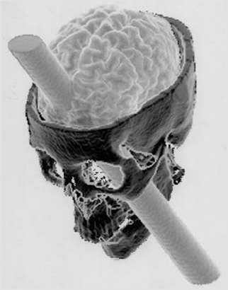
Figure 1.8: Computer reconstruction
scien tific knowledge
of the skull of Phineas Gage illustrating
occur as small
the projection of the tamp ing rod through
modifications in
the brain. Reprinted with permis sion
extant knowledge.
from Damasio, H., et al. 1994. The
return of Phineas Gage: Clues about the
brain from a famous patient. Science
264:1102-05.
Sample Answers to Questions on Master 1.6
Question 1. How did Phineas Gage change after the accident?
After the accident, Gage’s personality changed. He was no longer the
like able and responsible person he was before the accident. Instead
he was irresponsible and used profanity.
Question 2. How did Phineas Gage’s accident change scientists’
under standing of the brain?
Scientists learned that the brain does more than control language
and movement. It also controls emotions and social behaviors.
Equally impor tant, scientists learned that the brain processes
information for specific functions in specific brain areas.
34
Activity 5: Where Do Drugs Act?
1. Now that students understand that different areas in the brain
process specific types of stimuli, ask students to consider things
Content Standard C:
that make them feel good, or are pleasurable. How might doing
something pleasurable change brain activity?
Multicellular organisms
have nervous systems
If students understand, from Activity 2 of this lesson, that brain
that generate behavior.
functions are localized to specific brain areas, they should suspect
Content Standard F:
that things that make them feel pleasure will stimulate a specific
An individual’s mood
brain region.
and behavior may be
2. Display the transparency of Master 1.7, The Reward System. Tell modified by substanc