Step 2
Activity 1. Have students identify the part of the brain that was
active in each case.
Activity 4: Who Was Phineas Gage?
Give each student a copy of Master 1.6, and ask them to read the
Page 34
story and answer the questions.
Step 1
Activity 5: Where Do Drugs Act?
Ask students to consider things that make them feel good or are
Page 35
pleasurable. Have them consider the question, How might doing
Step 1
something pleasurable change brain activity?
Display a transparency of Master 1.7. Tell students that part of
Page 35
the brain produces and regulates feelings of pleasure, which
Step 2
scientists call reward. Point out the parts of the brain that make up
the reward system: the ventral tegmental area (VTA), the nucleus
accumbens, and part of the frontal region of the cerebral cortex.
Introduce students to the idea that drugs of abuse activate the
Page 35
brain’s reward system. Specifically, introduce the idea that the
Step 3
action of drugs on the reward center is what makes the user feel
pleasure and want to continue taking drugs.
Ask students to hypothesize how PET images of a person’s brain
Page 35
would change after taking drugs of abuse. Inform them that
Step 4
they will learn more about how drugs affect the brain during the
remaining lessons in this unit.
= Involves copying a master.
= Involves making a transparency.
39
Student Lesson 1
L E S S O N 2
Explore/Explain
Neurons, Brain
Chemistry, and
Neurotransmission
Source: NIDA. 1996. The Brain & the Actions of Cocaine,
Opiates, and Marijuana. Slide Teaching Packet for Scientists.
Overview
At a Glance
Students learn that the neuron is the functional unit of the brain. To
learn how neurons convey information, students analyze a sequence of
illustrations and watch an animation. They see that neurons communicate
using electrical sig nals and chemical messengers called neurotransmitters
that either stimulate or inhibit the activity of a responding neuron. Students
then use the informa tion they have gained to deduce how one neuron
influences the action of another.
Major Concept
Neurons convey information using electrical and chemical signals.
Objectives
By the end of these activities, the students will
• understand the hierarchical organization of the brain, neuron,
and synapse;
• understand the sequence of events involved in communication
at the synapse; and
• understand that synaptic transmission involves neurotransmitters
that may be either excitatory or inhibitory.
Basic Science–Health Connection
Communication between neurons is the foundation for brain function.
Under standing how neurotransmission occurs is crucial to understanding
how the brain processes and integrates information. Interruption of neural
communi cation causes changes in cognitive processes and behavior.
41
The Brain: Understanding Neurobiology Through the Study of Addiction
The Brain Is Made Up of Nerve Cells and Glial Cells
Background
The brain of an adult human weighs about 3 pounds and contains billions
Information
of cells. The two distinct classes of cells in the nervous system are neurons
(nerve cells) and glia (glial cells).
The basic signaling unit of the nervous system is the neuron. The brain
contains billions of neurons; the best estimates are that the adult human
brain contains 1011 neurons. The interactions between neurons enable
people to think, move, main tain homeostasis, and feel emotions. A neuron
is a specialized cell that can pro duce different actions because of its precise
connections with other neurons, sensory receptors, and muscle cells. A
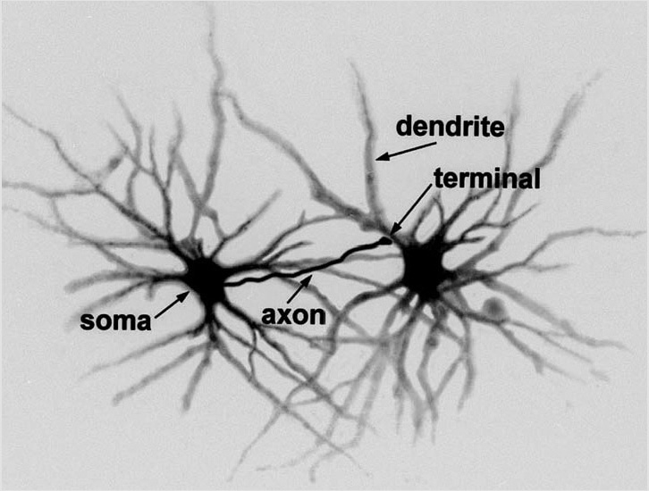
typical neuron has four morphologically defined regions: the cell body,
dendrites, axons, and presynaptic, or axon, terminals.1,2,3
Figure 2.1: The neuron, or nerve cell, is the functional unit of the nervous system.
The neuron has processes called dendrites that receive signals and an axon that
transmits signals to another neuron.
The cell body, also called the soma, is the metabolic center of the neuron.
The nucleus is located in the cell body, and most of the cell’s protein
synthesis occurs in the cell body.
A neuron usually has multiple processes, or fibers, called dendrites that
extend from the cell body. These processes usually branch out somewhat
like tree branches and serve as the main apparatus for receiving input into
the neuron from other nerve cells.
The cell body also gives rise to the axon. Axons can be very long processes;
in some cases, they may be up to 1 meter long. The axon is the part of the
neuron that is specialized to carry messages away from the cell body and
to relay messages to other cells. Some large axons are surrounded by a fatty
insulating material called myelin, which enables the electrical signals to
travel down the axon at higher speeds.
Near its end, the axon divides into many fine branches that have specialized
swellings called axon, or presynaptic, terminals. These presynaptic
terminals end in close proximity to the dendrites of another neuron. The
dendrite of one neuron receives the message sent from the presynaptic
terminal of another neuron.
42
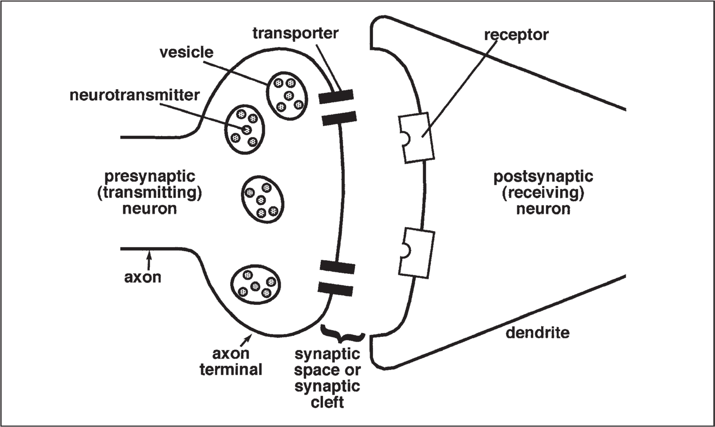
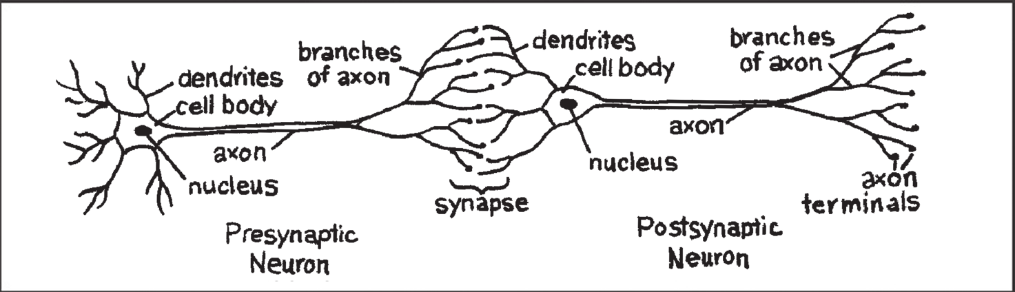
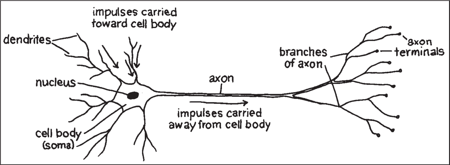
Figure 2.2: Neurons transmit information to other neurons. Information passes from
the axon of the presynaptic neuron to the dendrites of the postsynaptic neuron.
The site where a presynaptic terminal ends in close proximity to a receiving
dendrite is called the synapse. The cell that sends out information is called
the presynaptic neuron, and the cell that receives the information is called
the postsynaptic neuron. It is important to note that the synapse is not
a physical con nection between the two neurons; there is no cytoplasmic
continuity between the two neurons. The intercellular space between the
presynaptic and postsy naptic neurons is called the synaptic space or
synaptic cleft. An average neu ron forms approximately 1,000 synapses
with other neurons. It has been estimated that there are more synapses in
the human brain than there are stars in our galaxy. Furthermore, synaptic
connections are not static. Neurons form new synapses or strengthen
synaptic connections in response to life experi ences. This dynamic change
in neuronal connections is the basis of learning.
Figure 2.3: The synapse is the site where chemical signals pass between neurons.
Neurotransmit ters are released from the presynaptic neuron terminals into the
extracellular space, the synaptic cleft or synaptic space. The released neurotransmitter
molecules can then bind to specific recep tors on the postsynaptic neuron to elicit
a response. Excess neurotransmitter can then be reabsorbed into the presynaptic
neuron through the action of specific reuptake molecules called transporters.
This process ensures that the signal is terminated when appropriate.
43
Student Lesson 2
The Brain: Understanding Neurobiology Through the Study of Addiction
The brain contains another class of cells called glia. There are as many
as 10 to 50 times more glial cells than neurons in the central nervous
system. Glial cells are categorized as microglia or macroglia. Microglia
are phago cytic cells that are mobilized after injury, infection, or disease.
They are derived from macrophages and are unrelated to other cell types
in the ner vous system. The three types of macroglia are oligodendrocytes,
astrocytes, and Schwann cells. The oligodendrocytes and Schwann cells
form the myelin sheaths that insulate axons and enhance conduction of
electrical signals along the axons.
Scientists know less about the functions of glial cells than they do about the
functions of neurons. Glial cells fulfill a variety of functions including as
• support elements in the nervous system, providing structure and
separating and insulating groups of neurons;
• oligodendrocytes in the central nervous system and Schwann cells
in the peripheral nervous system, which form myelin, the sheath that
wraps around cer tain axons;
• scavengers that remove debris after injury or neuronal death;
• helpers in regulating the potassium ion (K+) concentration in the
extracel lular space and taking up and removing chemical neurotrans-
mitters from the extracellular space after synaptic transmission;
• guides for the migration of neurons and for the outgrowth of axons
during development; and
• inducers of the formation of impermeable tight junctions in endo-
thelial cells that line the capillaries and venules of the brain to form
the blood-brain barrier.3
The Blood-Brain Barrier
The blood-brain barrier protects the neurons and glial cells in the brain from substances that
could harm them. Endothelial cells that form the capillaries and venules make this barrier, forming
impermeable tight junc tions. Astrocytes surround the endothelial cells and induce them to form
these junctions. Unlike blood vessels in other parts of the body that are relatively leaky to a variety
of molecules, the blood-brain barrier keeps many substances, including toxins, away from the
neurons and glia.
Most drugs do not get into the brain. Only drugs that are fat soluble can penetrate the blood-brain
barrier. These include drugs of abuse as well as drugs that treat mental and neurological illness.
The blood-brain barrier is important for maintaining the environment of neurons in the brain, but it
also presents challenges for scientists who are investigating new treatments for brain disorders. If a
medication cannot get into the brain, it cannot be effective. Researchers attempt to circumvent the
problems in different ways. Some techniques alter the structure of the drug to make it more lipid
soluble. Other strategies attach potential therapeutic agents to molecules that pass through the
blood-brain bar rier, while others attempt to open the blood-brain barrier.4
44
Neurons Use Electrical and Chemical Signals to
Transmit Information*
The billions of neurons that make up the brain coordinate thought, behavior,
homeostasis, and more. How do all these neurons pass and receive information?
Neurons convey information by transmitting messages to other neurons
or other types of cells, such as muscles. The following discussion focuses
on how one neuron communicates with another neuron. Neurons employ
electrical signals to relay information from one part of the neuron to
another. The neu ron converts the electrical signal to a chemical signal in
order to pass the information to another neuron. The target neuron then
converts the message back to an electrical impulse to continue the process.
Within a single neuron, information is conducted via electrical signaling.
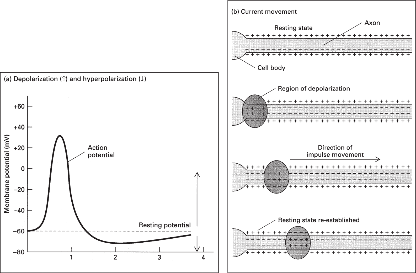
When a neuron is stimulated, an electrical impulse, called an action
potential, moves along the neuron axon.5 Action potentials enable signals
to travel very rapidly along the neuron fiber. Action potentials last less
than 2 milliseconds (1 millisecond = 0.001 second), and the fastest action
potentials can travel the length of a football field in 1 second. Action
potentials result from the flow of ions across the neuronal cell membrane.
Neurons, like all cells, maintain a balance of ions inside the cell that differs
from the balance outside the cell. This uneven distribution of ions creates
an electrical poten tial across the cell membrane. This is called the resting
membrane potential. In humans, the resting membrane potential ranges
from –40 millivolts (mV) to –80 mV, with –65 mV as an average resting
membrane potential. The resting membrane potential is, by convention,
assigned a negative number because the inside of the neuron is more negatively
charged than the outside of the neuron. This negative charge results from
the unequal distribu tion of sodium ions (Na+), potassium ions (K+), chloride
ions (Cl–), and other organic ions. The resting membrane potential is
maintained by an energy-dependent Na+-K+ pump that keeps Na+ levels
low inside the neuron and K+ levels high inside the neuron. In addition,
the neuronal membrane is more permeable to K+ than it is to Na+, so K+
tends to leak out of the cell more readily than Na+ diffuses into the cell.
A stimulus occurring at the cell body starts an electrical change that travels
like a wave over the length of the neuron. This electrical change, the action
potential, results from a change in the permeability of the neuronal membrane.
Sodium ions rush into the neuron, and the inside of the cell becomes more
positive. The Na+-K+ pump then restores the balance of sodium and potassium
to resting levels. However, the influx of Na+ ions in one area of the neuron
fiber starts a similar change in the adjoining segment, and the impulse
moves from the cell body toward the axon terminal. Action potentials are
an all-or-none phenomenon. Regardless of the stimuli, the amplitude and
duration of an action potential are the same. The action poten tial either
occurs or it doesn’t. The response of the neuron to an action poten tial
depends on how many action potentials it transmits and their frequency.
* “Electrical signals” are not actually electric because ions travel down the axon, not
electrons. For the sake of simplicity, though, we use “electrical.”
45
Student Lesson 2
The Brain: Understanding Neurobiology Through the Study of Addiction
Figure 2.4: (a) Recording of an action potential in an axon following stimulation due to changes in the permeability of the cell membrane to sodium and potassium ions. (b) The cell membrane of a resting neuron is more negative on the inside of the cell than on the outside. When the neuron is stimulated, the permeability of the membrane changes, allowing Na+ to rush into the cell. This causes the inside of the cell to become more positive. This local change starts a similar change in the adjoining segment of the neuron’s membrane. In this manner, the electrical impulse moves along the neuron. From: Molec ular Cell Biology , by Lodish et al. 1986, 1990 by Scientific American Books, Inc. Used with permission by W.H. Freeman and Company.
Electrical signals carry information within a single neuron. Communication
between neurons (with a few exceptions in mammals) is a chemical process.
When the neuron is stimulated, the electrical signal (action potential)
travels down the axon to the axon terminals. When the electrical signal
reaches the end of the axon, it triggers a series of chemical changes in the
axon terminal. Cal cium ions (Ca++) flow into the axon terminal, which
then initiates the release of neurotransmitters. A neurotransmitter is a
molecule that is released from a neuron to relay information to another cell.
Neurotransmitter molecules are stored in membranous sacs called vesicles
in the axon terminal. Each vesicle contains thousands of molecules of a
given neuro transmitter. For neurons to release their neurotransmitter, the
vesicles fuse with the neuronal membrane and then release their contents,
the neurotrans mitter, via exocytosis. The neurotransmitter molecules are
released into the synaptic space and diffuse across the synaptic space to
the postsynaptic neu ron. A neurotransmitter molecule can then bind to a
special receptor on the membrane of the postsynaptic neuron. Receptors
are membrane proteins that are able to bind a specific chemical substance,
46
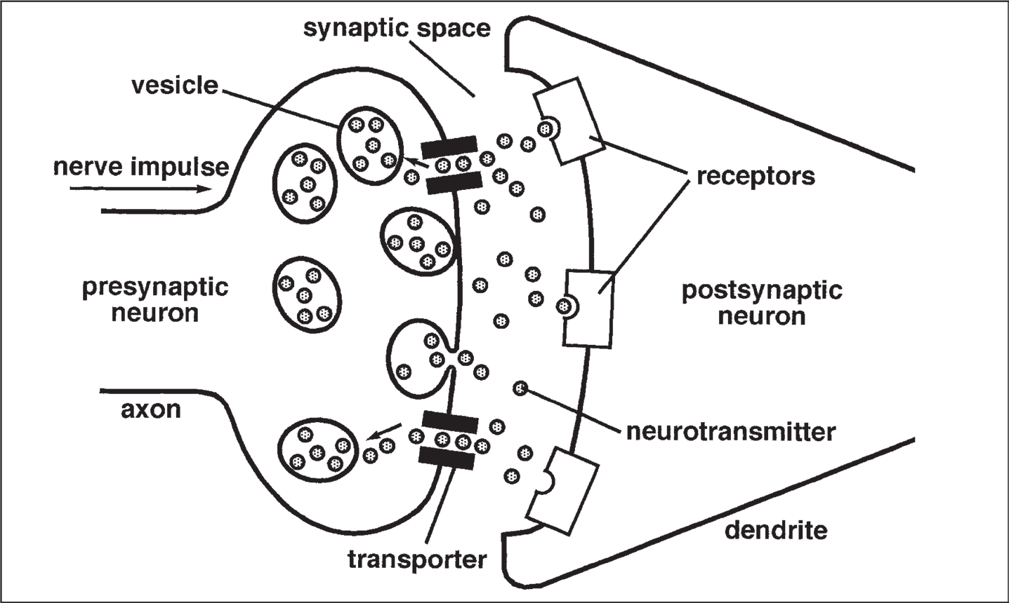
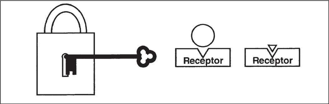
Figure 2.5: Schematic diagram of a synapse. In response to an electrical impulse,
neuro transmitter molecules released from the presynaptic axon terminal bind to the
specific receptors for that neurotransmitter on the postsynaptic neuron. After binding
to the recep tor, the neurotransmitter molecules either may be taken back up into the
presynaptic neu ron through the transporter molecules for repackaging into vesicles or
may be degraded by enzymes present in the synaptic space.
such as a neurotransmitter. For example, the dopamine receptor binds the
neurotransmitter dopamine but does not bind other neurotransmitters such
as serotonin. The interaction of a receptor and neurotransmitter can be
thought of as a lock-and-key for regulat ing neuronal function. Just as a key
fits only a specific lock, a neurotransmit ter only binds with high affinity to
a specific receptor. The chemical binding of neurotransmitter and receptor
initiates changes in the postsynaptic neuron that may facilitate or inhibit
an action potential in the postsynaptic neuron. If it does trigger an action
potential, the communication process continues.
Figure 2.6: Like a lock that will open only if the right key is used, a receptor will
bind only a molecule that has the right chemical shape. Molecules that do not have
the right “fit” will not bind to the receptor and will not cause a response.
After a neurotransmitter molecule binds to its receptor on the postsynaptic
neuron, it comes off (is released from) the receptor and diffuses back
into the synaptic space. The released neurotransmitter, as well as any
neurotransmitter that did not bind to a receptor, is either degraded by
enzymes in the synaptic cleft or taken back up into the presynaptic
axon terminal by active transport through a transporter or reuptake
47
Student Lesson 2
The Brain: Understanding Neurobiology Through the Study of Addiction
pump. Once the neurotransmitter is back inside the axon terminal, it is
either destroyed or repackaged into new vesicles that may be released
the next time an electrical impulse reaches the axon terminal. Different
neurotransmit ters are inactivated in different ways.
Neurotransmitters Can Be Excitatory or Inhibitory
Different neurotransmitters fulfill different functions in the brain.
Some neu rotransmitters act to stimulate the firing of a postsynaptic
neuron. Neuro transmitters that act this way are called excitatory
neurotransmitters because they lead to changes that generate an action
potential in the responding neu ron.1,6 Other neurotransmitters, called
inhibitory neurotransmitters, tend to block the changes that cause an
action potential to be generated in the responding cell. Table 2.1 lists
some of the “classical neurotransmitters” used in the body and their
major functions. In addition to the so-called classical neurotransmitters,
there are many other peptide transmitters, sometimes called neuromodulators.
They are similar to classical neurotransmitters in the way they are stored
(in vesicles) and released, but they differ in how they are inactivated.
Most neurons contain multiple transmitters, often a classical one (such as
dopamine) and one or more peptides (such as neurotensin or endorphins).
The postsynaptic neuron often receives and integrates both excitatory
and inhibitory mes sages. The response of the postsynaptic cell depends
on which message is stronger. Keep in mind that a single neurotransmitter
molecule cannot cause an action potential in the responding neuron. An
action potential occurs when many neurotransmitter molecules bind to
and activate their receptors. Each interaction contributes to the membrane
permeability changes that generate the resultant action potential.
Table 2.1: Major Neurotransitters in the Body1,6,7
Neurotransmitter
role in the body
Acetylcholine
Used by spinal cord motor neurons to cause muscle contraction
and by many neurons in the brain to regulate memory. In most
instances, acetylcholine is excitatory.
Dopamine
Produces feelings of pleasure when released by the brain reward
system. Dopamine has multiple functions depending on where in
the brain it acts. It is usually inhibitory.
GABA (gamma-aminobutyric acid) The major inhibitory neurotransmitter in the brain. It is important
in producing sleep, reducing anxiety, and forming memories.
Glutamate
The most common excitatory neurotransmitter in the brain. It is
important in learning and memory.
Glycine
Used mainly by neurons in the spinal cord. It probably always acts
as an inhibitory neurotransmitter.
Norepinephrine
Acts as a neurotransmitter and a hormone. In the peripheral
ner vous system, it is part of the fight-or-flight response. In the
brain, it acts as a neurotransmitter regulating blood pressure and
calmness. Norepinephrine is usually excitatory, but it is inhibitory
in a few brain areas.
Serotonin
Involved in many functions including mood, appetite, and sensory
perception. In the spinal cord, serotonin is inhibitory in pain pathways.
48
Web-based Activities
In Advance
Activity
Web Component?
1
No
2
Yes
3
Yes
4
Yes
Photocopies
For each group
For the class
For each student
of 3 students
1 transparency of Master 2.1, Anatomy of a Neuron
1 copy of Master 2.3, 1 copy of Master 2.5,
1 transparency of Master 2.2, Neurons Interact with
Other Neurons Through Synapses
1 copy of Master 2.7,
1 transparency of Master 2.4,