Different clinicians take different numbers of clinical photographs, depending on who you talk to! There is no “standard” set that is universally-approved as a rule of thumb. However, it can generally be accepted - based on many authorities’ opinions in this field - that a complete “Clinical Photographic Set” for any orthodontic patient at any stage of treatment, that would enable the clinician to obtain maximum benefit and information, should include a minimum of NINE photographs; FOUR extra-oral, and FIVE intraoral photographs.
Extra-oral clinical photographs are the easiest photographs to take. They only require proper positioning of the patient and clinician, in addition of course to the digital camera setup itself. Intra-oral photos require - in addition to the camera setup - the proper cheek retractors, dental photography mirrors, as well as a well-trained assistant if possible. Clinical steps for properly taking each photograph is explained further.

Extra-oral photos consist of the following four shots:
1. Face-Frontal (lips relaxed).
2. Face-Frontal (Smiling).
3. Profile (Right side preferably - Lips relaxed).
4. (45 °) Profile (also known as 3/4 Profile - Smiling).
Face - Frontal (Lips Relaxed)

The first extra-oral photo to be usually taken, this photo is probably the easiest. However, there are still some important guidelines that need to be taken into account when taking this shot.
First, the Framing of the shot, should encompass the whole of the patient’s face and neck, with a reasonable margin of space all around. This is ensured by holding the camera lens in a vertical position, and by standing a reasonable distance away from the patient when
taking the shot. The following general guidelines should also be noted:A. The patient should stand with their head in the Natural Head Position, with eyes looking straight into the camera lens.
B.The patient should hold their teeth and jaw in a relaxed (Rest) position, with the lips in contact (if possible) and in a relaxed position.
C.Make sure patient’s head is not tilted or their face rotated to either side; the shot should be taken at 90° to the facial mid-line from the front.
D. Ensuring the patient’s inter-pupillary line is leveled is very important.
It is recommended that the patient stands in front of a plain, dark or whitecolored wall or background when taking all extra-oral shots. This is to ensure maximum clarity of facial features and outlines without the presence of distracting objects in the background.
 Framing
 Face - Frontal (Smiling)

The same guidelines as for the Face - Frontal shot apply here, with the simple but important exception that the patient should be SMILING in a natural way, with the teeth visible. This photo greatly aids in visualizing the patient’s Smile esthetics and soft tissue proportions during smiling.
Profile (Right Side - Lips Relaxed)The Profile photo has a high diagnostic value to the orthodontist. After taking the frontal face photos, the patient is asked to bodily turn to their left, thus having their right profile side facing the clinician. The head should be in the Natural Head Position, with their eyes

fixed horizontally (preferably at a specific point at eye-level, or at the reflection of their own pupils in a mirror). The wrong head posture can result in confusion regarding the patient’s actual skeletal pattern.
Ideally, the whole of the right side of the face should be clearly visible with no obstructions such as hair, hats or scarfs.
For the most useful, professional-looking photo possible, the use of the RingFlash is essential. As explained earlier, the Ring-Flash will eliminate any shadowing of the border of the patient’s profile onto the background, which can compromise the quality of the photo considerably.

The final extra-oral photograph to be taken, this shot conveys the patient as if in “social interaction”, and can give valuable information about the smile esthetics’ changes pre- and post-treatment.
From the Profile photo position, the patient is asked to turn their heads slightly to their right (about 3/4 of the way - hence the name), while keeping their body still in the previous “Profile Shot” position i.e. Facing forward. They are then instructed to look into the camera mostly by turning their eyes further to the right
to meet the lens, and then smile. It is essential that the patient’s teeth show clearly when smiling, otherwise the photograph would be of minimum benefit.• The background used in taking the photos should be either a solid-white background (or a back-lit light-box), or a solid-dark color such as Dark Blue. Taking extra-oral photos with the patient sitting on the dental chair or with multiple objects in the background should be avoided.
• The clinician’s positioning for these photos would be standing a few feet away from the patient, and at the same eye level if possible. Younger and shorter patients can stand on a special stand to get them to reach a suitable height if needed.
• All extra-oral photos require that the Aperture value (F value) be set to a minimum e.g F8 is usually a suitable setting.

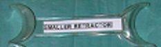
Intra-oral photographs require more attention to detail to produce good results. For these shots, the use of the special cheek retractors and dental mirrors will be required, in addition to help from a dental assistant.
There are five required intra-oral photographs:
1. Frontal - in occlusion
2. Right Buccal - in occlusion
3. Left Buccal - in occlusion
4. Upper Occlusal
5. Lower Occlusal
Frontal - in Occlusion
The first photo usually to be taken of the set. With the patient sitting comfortably in the dental chair and raised to elbow-level of the clinician, the assistant
patient and uses the
stands behind the first larger set of
retractors from the wide ends to retract the patient’s lips sideways and away from the

teeth and gingivae , towards the clinician. This is important to allow maximum visualization of all teeth and alveolar ridges in the photograph, and also to minimize any discomfort for the patient from retractor edges impinging on the


gingivae. The photo should be taken 90° to the facial mid-line using the upper frenal attachment as a guide. The dental mid-lines are not as reliable for this purpose as they can be shifted to one side or the other depending on the malocclusion present. The full extension of the sulci is paramount for full visualization and clarity, and the high F value setting e.g. F32 is required to attain maximum depth of field of the shot with even the last visible molars fully in focus. The Ring-Flash will greatly aid in producing a quality photograph by ensuring the best possible light distribution of the image without shadows, especially of the deeper parts of the oral cavity and buccal vestibules.
Right Buccal - in Occlusion
Usually the second shot in the series. The assistant flips the right retractor to the narrower side, while the left retractor remains in place as for the previous frontal shot. The patient is asked to turn their head slightly to their left so their right side will be facing the clinician. Here, the clinician holds the right

retractor and stretches it to the extent that the last present molar is visible if possible, while the assistant maintains hold of the left retractor, without undue
stretching. Again, the shot is taken 90° to the canine-premolar area for best visualization of the buccal segment relationship, as this is very important in orthodontic assessment. A useful tip would be to for the clinician to fully stretch the right retractor just before taking the shot to minimize any discomfort for the patient, and achieve maximum visibility of the last molar.
Left Buccal - in Occlusion
The third shot in the series, it is very similar to the Right Buccal shot. The assistant now switches the retractors with the narrow end on the photo side (patient’s left) and the wide end on the other (patient’s right). Again, the shot is taken at 90° to the canine-premolar area, and to ensure this, the clinician should

move their body slightly to the right while holding the retractor on the photo side, while the patient turns their head slightly to their right.


Upper Occlusal - Mirror
Here, the dental mirrors come into play. The assistant now switches to the smaller retractor set and with the patient’s mouth held open, the retractors are inserted in a “V” shape to retract the upper lips sideways and away from the teeth. The clinician inserts the mirror with its wider end inwards to capture maximum width of the arch posteriorly, and pull it slightly downwards so that the whole upper arch is visible to the last present molar. The patient may be instructed to lower their head slightly so that the shot can be taken 90° to the plane of the mirror for best visibility. It is recommended that the mid-palatal raphe is used as a guide for the orientation of the shot to get it leveled. Minimum retractor show in the image is

recommended, and no fingers should be visible at any time.
Lower Occlusal - Mirror
The final shot in the series. The assistant would now lower the smaller retractors into a Reverse “V” shape to retract the lower lips sideways and away form the teeth. The clinician would now lift the mirror upwards so he/she may visualize

“Ideal” Shot - Tongue Rolled Back

“Less-than-Ideal” Shot - Tongue Visible But Not Obstructing View
the reflection of the lower arch, while the patient is be asked to “lift their chin up” slightly. Ideally, the shot should be taken 90° to the plane of the mirror, with the last molar present visible. An important issue here would be the tongue position of the patient while taking the photo. It is best to ask the patient to “roll back”
their tongue behind the mirror so that it won’t interfere with the visibility of any teeth, particularly in the posterior area.
The following images reveal in a more visual way some important aspects of Clinician/Assistant positioning, as well as retraction technique during photographic record-taking.
 Upper Occlusal Shot
 Lower Occlusal Shot
 Position Of Retractors For Upper Occlusal Shot

Position Of Retractors For Lower Occlusal Shot

Right Buccal Shot:
Clinician Holding Narrow-End Retractor On Side To Be
 Helpful Tips

•The direction of pull of the retractors is always sideways and slightly forward, away from the gingival tissues. This maximizes the field of view and minimizes patient discomfort.
•Wetting the retractors just before insertion eases the process of positioning them properly with minimum patient discomfort.

•When taking occlusal “Mirror” shots, slightly warming the mirror in warm water prior to insertion helps prevent “Fogging” of the mirrors which would prevent a clear image.
•In certain cases, profuse salivary flow and “frothing” can affect the quality of the image being taken, thus a saliva ejector can be used to eliminate saliva prior to taking each photograph.
•During occlusal “mirror” shots, instruct the patient to “open wide” just prior to pressing the camera button. This helps in obtaining the maximum mouth opening at the right moment, and minimizes the patient’s fatigue during the procedure.
•It is recommended that all photographic records be taken before impressiontaking, to eliminate the possibility of impression material being stuck between the teeth or the face during photographic record-taking.